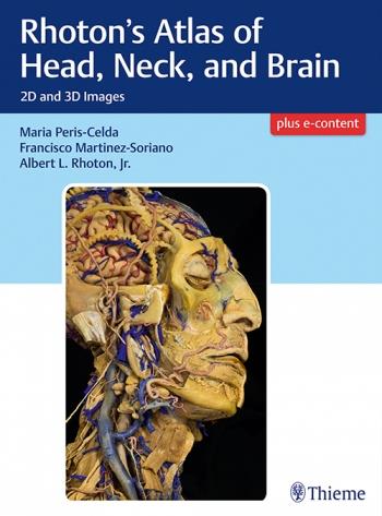
Rhoton`s Atlas of Head, Neck, and Brain, 2D and 3D Images
ΑΣΦΑΛΗ ΠΛΗΡΩΜΗ
ΑΠΟΣΤΟΛΗ
ΔΩΡΕΑΝ ΕΠΙΣΤΡΟΦΗ
Λεπτομέρειες
Masterful 2D and 3D head, neck, and brain dissections provide unsurpassed insights into head, neck, and brain anatomy
An internationally renowned and beloved author, educator, brain anatomist, and neurosurgeon, Professor Albert Rhoton has a special place in medical history. He was revered by students and colleagues and is regarded as one of the fathers of modern microscopic neurosurgery. A driving principle in his anatomy lab was the simple phrase, \"Every Second.\" This was embraced in his philosophy that every second of every day, a patient\'s life was improved by a surgeon assisted by the anatomic knowledge his lab helped elucidate and distribute.
Rhoton\'s Atlas of Head, Neck, and Brain is the visually exquisite crowning achievement of Dr. Rhoton\'s brilliant career and unwavering dedication to the intertwined pursuits of surgical anatomy and neurosurgery. The atlas reflects the unparalleled contributions Dr. Rhoton made to the contemporary understanding of neurosurgical anatomy. Dr. Peris-Celda, with the collaboration of an impressive cadre of international multidisciplinary experts, worked closely under Dr. Rhoton\'s tutelage on this project. This book is the culmination of 5 years of work and experience gleaned from more than 40 years of surgical anatomy research and exquisite dissection techniques performed in Dr. Rhoton\'s laboratory.
Special Features
- Each anatomic dissection meticulously labeled with English and Latin descriptors for easy cross referencing with other resources.
- Multiple views of the most complex regions of the head, neck, and brain provide a deeper understanding of anatomy.
- More than 600 anatomical images systematically organized in four major sections: Osteology of the Head and Neck; Face and Neck; Ear, Nose, Pharynx,
- Larynx, and Orbit; and Neuroanatomy and Cranial Base.
- Superb 2D images presented in a large printed format to optimize the viewing experience.
- 3D digital images fully realize the beauty of the dissections and enhance the learning process.
- Specimens injected with colored silicone provide better visualization of arteries and veins.
Breathtakingly stunning, this atlas is certain to be a treasured reference for medical students, residents, and clinicians specializing in neurosurgery, facial plastic surgery, otolaryngology, maxillofacial surgery, and craniofacial surgery for many years to come.
- Μεταφορές - Επιστροφές
- Αρχεία
Περιγραφή Προϊόντος
Masterful 2D and 3D head, neck, and brain dissections provide unsurpassed insights into head, neck, and brain anatomy
An internationally renowned and beloved author, educator, brain anatomist, and neurosurgeon, Professor Albert Rhoton has a special place in medical history. He was revered by students and colleagues and is regarded as one of the fathers of modern microscopic neurosurgery. A driving principle in his anatomy lab was the simple phrase, \"Every Second.\" This was embraced in his philosophy that every second of every day, a patient\'s life was improved by a surgeon assisted by the anatomic knowledge his lab helped elucidate and distribute.
Rhoton\'s Atlas of Head, Neck, and Brain is the visually exquisite crowning achievement of Dr. Rhoton\'s brilliant career and unwavering dedication to the intertwined pursuits of surgical anatomy and neurosurgery. The atlas reflects the unparalleled contributions Dr. Rhoton made to the contemporary understanding of neurosurgical anatomy. Dr. Peris-Celda, with the collaboration of an impressive cadre of international multidisciplinary experts, worked closely under Dr. Rhoton\'s tutelage on this project. This book is the culmination of 5 years of work and experience gleaned from more than 40 years of surgical anatomy research and exquisite dissection techniques performed in Dr. Rhoton\'s laboratory.
Special Features
- Each anatomic dissection meticulously labeled with English and Latin descriptors for easy cross referencing with other resources.
- Multiple views of the most complex regions of the head, neck, and brain provide a deeper understanding of anatomy.
- More than 600 anatomical images systematically organized in four major sections: Osteology of the Head and Neck; Face and Neck; Ear, Nose, Pharynx,
- Larynx, and Orbit; and Neuroanatomy and Cranial Base.
- Superb 2D images presented in a large printed format to optimize the viewing experience.
- 3D digital images fully realize the beauty of the dissections and enhance the learning process.
- Specimens injected with colored silicone provide better visualization of arteries and veins.
Breathtakingly stunning, this atlas is certain to be a treasured reference for medical students, residents, and clinicians specializing in neurosurgery, facial plastic surgery, otolaryngology, maxillofacial surgery, and craniofacial surgery for many years to come.


test
Μεταφορές - Επιστροφές
Επιστροφές
ΠΟΛΙΤΙΚΗ ΕΠΙΣΤΡΟΦΩΝ
Στόχος μας είναι να εξασφαλίσουμε την απόλυτη ικανοποίησή σας. Αν για οποιονδήποτε λόγω, δεν μείνετε ευχαριστημένοι από την παραγγελία σας, μπορείτε να ασκήσετε το δικαίωμά σας να επιστρέψετε μέρος ή όλα τα προϊόντα που αγοράσατε εντός δεκατεσσάρων (14) ημερών από την ημερομηνία παραλαβής. Αν δεν υπάρχει διαθέσιμο
το ανάλογο προϊόν που επιθυμείτε, το ποσό διαφοράς που προκύπτει διατηρείται στον λογαριασμό σας για να το χρησιμοποιήσετε για μία άλλη αγορά. Θα γίνεται αποδεκτή μόνο η επιστροφή των προϊόντων που βρίσκονται σε άριστη κατάσταση. Παρακαλώ, σημειώστε ότι η αποστολή του μη επιθυμητού προϊόντος γίνεται με εταιρεία courier με χρέωση της εταιρίας μας. Η αποστολή του νέου προϊόντος γίνεται μέσω εταιρεία courier με χρέωση πελάτη.
ΕΛΛΑΤΩΜΑΤΙΚΟ ΠΡΟΙΟΝ
Στην περίπτωση που λάβετε ένα ελαττωματικό ή κατεστραμμένο προϊόν το βιβλιοπωλείο μας θα καταβάλλει κάθε προσπάθεια για να αντικατασταθεί με ένα ίδιο ή παρόμοιο προϊόν χωρίς κάποιο επιπλέον κόστος για σας.
Παρακαλούμε επικοινωνήστε μαζί μας με e-mail στο ipokratis@ipokratis.gr ή στο 2310212212 για περαιτέρω διευκρινήσεις.